What Diagnostic Medical Sonographers Do
About this section
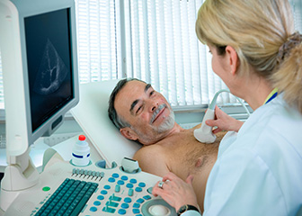
Diagnostic medical sonographers use high-frequency sound waves to produce images of the inside of the body.
Diagnostic medical sonographers, also called ultrasound technicians, operate special equipment to create images of inside the body. They work closely with physicians and surgeons, who view the images to assess and diagnose medical conditions.
Duties
Diagnostic medical sonographers typically do the following:
- Prepare patients by explaining the procedure to them and answering their questions
- Prepare exam rooms and maintain diagnostic imaging equipment
- Properly position patients for imaging
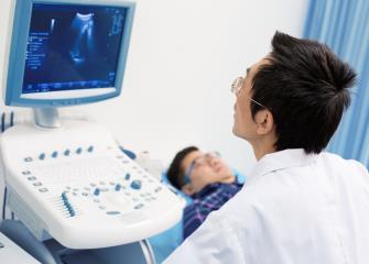
- Operate equipment to obtain diagnostic images
- Review images to check for quality and adequate coverage of the areas needed for diagnoses
- Analyze results for abnormalities and other diagnostic information and provide a summary of findings to physicians
- Record findings and keep track of patients’ records
Diagnostic medical sonographers specialize in creating images, known as sonograms or ultrasounds, that depict the body’s organs and tissues. Sonography is often the first imaging test performed when disease is suspected.
Sonography uses high-energy sound waves to produce images of the inside of the body. The sonographer uses an instrument called a transducer to scan parts of the patient’s body that are being examined. The transducer emits pulses of sound that bounce back, causing echoes. The echoes form an image on a computer that physicians use for diagnosis.
The following are types of diagnostic medical sonographers:
Abdominal sonographers specialize in imaging a patient’s abdominal cavity and nearby organs, such as the kidney, liver, gallbladder, pancreas, and spleen. Abdominal sonographers may assist with biopsies or other examinations requiring ultrasound guidance.
Breast sonographers specialize in imaging a patient’s breast tissue. Sonography can confirm the presence of cysts and tumors that may have been detected by the patient, the physician, or a mammogram. Breast sonographers assist with procedures that track tumors and help to provide information that will aid physicians in making decisions about treatment options for breast cancer patients.
Cardiac sonographers (echocardiographers) specialize in imaging a patient’s heart. They use ultrasound equipment to examine the heart’s chambers, valves, and vessels. An echocardiogram may be performed either while the patient is resting or after the patient has been physically active. Cardiac sonographers also may take echocardiograms of fetal hearts so that physicians can diagnose cardiac conditions during pregnancy.
Musculoskeletal sonographers specialize in imaging muscles, ligaments, tendons, and joints. These sonographers may assist with ultrasound guidance for injections, or during surgical procedures, that deliver medication or treatment directly to affected tissues.
Pediatric sonographers specialize in imaging of children and infants. Many of the medical conditions they image are associated with premature births or birth defects. Pediatric sonographers may work closely with pediatricians and other caregivers.
Obstetric and gynecologic sonographers specialize in imaging the female reproductive system. For example, many pregnant women receive sonograms to track the baby’s growth and health.
Vascular technologists (vascular sonographers) create images of blood vessels and collect data that help physicians diagnose disorders affecting blood flow. Vascular technologists often evaluate blood flow and identify blocked arteries or blood clots.